JAPAN/FUKUI
Department of Forensic Medicine, Graduate School of Medicine, The University of Tokyo
the Institute of Dinosaur Research, Fukui Prefectural University.
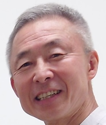
This summary recounts the evolution of my career and my involvement in postmortem imaging. In 1991, after graduating from university, I began working at a university hospital and a cancer center, where I spent 8 years. Seven of those 8 years were dedicated to my role as a radiologist and respiratory physician, engaging in imaging diagnosis (CT, MRI, ultrasound, bronchoscopy), treatment (chemotherapy, radiation therapy), and terminal care for lung cancer patients. Whenever a hospitalized patient passed away, I would recommend a pathological autopsy to the family. This practice allowed me to frequently compare antemortem (living) imaging findings with autopsy findings (radiologic-pathologic correlation). One of those 8 years I spent as an anesthesiologist in the operating room and conducted cardiopulmonary resuscitation managements in the emergency room (ER). What I learned regarding physiology, biochemistry, and pharmacology through my work experience in the fields of pulmonology and anesthesiology, not only anatomical and pathological studies as a radiologist, became the base knowledge for me in writing later research papers.
In 1999, I was transferred to Tsukuba Medical Center Hospital (TMCH), an emergency hospital where postmortem CT (PMCT) has been systematically performed for the first time in Japan since 1985 when the hospital was established. The purpose of the PMCT was to screen for causes of death in patients who were brought to the ER in cardiopulmonary arrest (CPA), were resuscitated, but subsequently died in the ER. (In Japan, the number of forensic pathologists is low, and the autopsy rate remains very limited.)
Upon my transfer to TMCH, I discovered a substantial archive of PMCT image data in the film reading room. Though emergency and critical care physicians referred to this valuable dataset as “a treasure trove”, they were too busy to analyze it. Therefore, I began analyzing the accumulated data and reported in 2000 that the PMCT findings could be classified into three main categories: cause of death, postmortem changes, and resuscitation-induced changes. At that time, I read Dr. Brogdon’s book “Forensic Radiology” published in 1998, which mainly focused on postmortem plain radiographs with limited descriptions regarding PMCT and postmortem MRI (PMMRI). TMCH also had an autopsy center within its facilities where forensic pathologists conducted autopsies on unusual deaths. I started to perform PMCT and PMMRI on bodies prior to autopsy, and presented the results of radiologic-pathologic correlation at conferences.
In 2006, a mystery novel involving postmortem imaging as a clue to solve problems became a bestselling novel in Japan; since then, the usefulness of postmortem imaging and the term ‘Autopsy imaging’ used in the novel have become widely known among public beyond medical professionals.
In 2015, I was transferred to my current hospital (equipped with 100 beds), where PMCT was already being performed on patients with CPA in the ER and sudden death cases among hospitalized patients. Currently, I am working with my role of in-hospital medical safety surveys, in addition to my primary role as a director of radiology department. I also I receive police inquests to inspect unusually deceased bodies. I perform PMCTs every few days in the evening between 17:00 and 24:00, and PMMRIs in some cases. The number of PMCTs has been especially increasing in Japan (60,000 cases in 2018 compared to 20,000 cases in 2012) though the autopsy rate has been remaining low. Further advancement in postmortem image interpretations, as well as distribution of the knowledge, will be of substantial help for death-cause detection in unusual deaths.
Name: Seiji Shiotani, MD, Ph.D
Date and place of birth: December 22, 1965, Kyoto City, Japan
Nationality: Japanese
Affiliation: Director of Radiology Department, Seirei Fuji General Hospital
3-1 Minami-cho, Fuji City, Shizuoka Prefecture 417-0026, Japan
e-mail: s.shiotani@sis.seirei.or.jp, Tel. +81-545-52-0780, Fax. +81-545-52-5837

Academic and occupational history:
Memberships:
Awards:
Forensic anthropology using CT in Japan
Suguru Torimitsu1,2
1 Department of Forensic Medicine, Graduate School of Medicine, The University of Tokyo
2 Education and Research Center of Legal Medicine, Graduate School of Medicine, Chiba University
Forensic anthropology is a branch of biological anthropology that is broadly defined as the scientific study of human skeletal remains, severely decomposed bodies, or body parts for identification. Identification of unknown remains is one of the most important aspects of medicolegal practice. A forensic anthropologist can estimate biological profiles such as ancestry, sex, age, and stature of unknown remains. Before the 1940s, the practice of forensic anthropology was limited to anatomists, physicians, and physical anthropologists. During this formative period, there was no formal instruction in the forensic applications of biological anthropology and little published research. From the 1940s to the early 1970s, attention from medicolegal and military agencies increased as they began to recognize the utility of forensic anthropology in the identification of deceased service members from WWII and the Korean War. Forensic anthropologists began using their expertise to assist law enforcement agencies in identifying victims of crimes. Forensic anthropology in Japan also has a long history; a lot of biological profile estimation methods using Japanese bones have been reported since the early 20th century. However, Japan did not have documented human skeletal collections. As a result, the sample size was small, and it was difficult to report our research internationally. Recently, postmortem CT scanning has become a useful tool for forensic practice and is routinely performed in some forensic departments. CT scanning can provide detailed information about the deceased. This information can be crucial for identifying victims, determining the cause of death, and reconstructing past events. CT has various advantages, such as being excellent at depicting bones, eliminating the need for maceration procedures, allowing data to be stored semi-permanently in little space, and being able to perform measurements even after autopsy. Therefore, CT images are useful for forensic anthropology research. Chiba University and the University of Tokyo introduced postmortem CT systems in 2009 and 2016, respectively, and we have a large amount of postmortem CT image data. We then performed various bone measurements and investigated their usefulness. During the lecture, some of our published research results and thoughts on the prospects of forensic anthropology will be presented.
Education
2015 Chiba University, PhD
2010 The University of Tokyo, Bachelor of Medicine
Employment & Work Experience
Department of Forensic Medicine, Graduate School of Medicine, The University of Tokyo
August 2024 – Current: Associate Professor
April 2015 – July 2024: Assistant Professor
Department of Legal Medicine, Graduate School of Medicine, Chiba University
September 2024 – Current: Project Associate Professor
April 2015 – August 2024: Project Assistant Professor
May 2012 – March 2015: Project Researcher
Centre for Forensic Anthropology, The University of Western Australia
May 2024 – Current: Adjunct Senior Lecturer
March 2023 – March 2024: Visiting research fellow
Tokyo Rosai Hospital
April 2010 – March 2012: Clinical Resident
Dinosaur Research in the Digital Age
The advent of digital technology, particularly X-ray CT scanners, has ushered in a new era in paleontology. These cutting-edge tools enable the non-destructive examination of fossilized remains, providing researchers with unprecedented insights into the internal structures of fossils. X-ray CT scanners, similar to those used in medical settings, have become indispensable in studying extinct vertebrates, including dinosaurs.
The application of X-ray CT scanning to paleontology began in the 1980s and has since evolved into a vital tool for examining the internal anatomy of fossils without causing damage. This technique is especially valuable in analyzing the cranial anatomy of dinosaurs, offering insights into the morphology of the brain, nerves, blood vessels, and inner ear structures.
In our research, we have utilized X-ray CT scanning to investigate the skull structures of various dinosaurs. For instance, we conducted a study on the lower jaw of Tyrannosaurus rex, where we discovered a complex network of trigeminal nerves, indicating a highly sensitive sense of touch in the jaw. This finding revealed that T. rex could use its jaw tips delicately despite its reputation as a powerful predator.
Additionally, we analyzed the well-preserved skull of Fukuiraptor, a theropod dinosaur discovered in Katsuyama, Fukui Prefecture, Japan. The X-ray CT scans of Fukuiraptor‘s skull revealed an exceptionally developed olfactory bulb and semicircular canals, indicating sharp senses of smell and balance. These findings suggest that Fukuiraptor was a highly agile animal, relying on its keen senses for survival.
The continuous unearthing of dinosaur fossils, combined with rapid advancements in CT scanning technology and image analysis software, promises to drive further breakthroughs in our understanding of dinosaur evolution and behavior. The integration of deep learning techniques in image analysis is poised to significantly enhance the accuracy and breadth of future research, opening up new vistas into the lives of these ancient creatures.
Soichiro Kawabe, PhD
Paleontologist.
Professor at the Institute of Dinosaur Research, Fukui Prefectural University.

Researcher at the Fukui Prefectural Dinosaur Museum.
Born in 1985. Completed a Ph.D. program at the Graduate School of Science, the University of Tokyo.
Specializes in the comparative morphology of vertebrates, with a particular focus on the brain morphology of dinosaurs, including birds, and mammals.